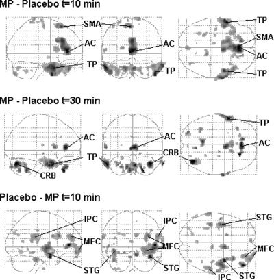
Figure 3.

Glass brains (sagittal, coronal, and transverse projections) showing the location of significant relative increases (MP‐placebo) and decreases (Placebo‐MP) in rCBF at 10 and 30 min after MP administration (P < 0.001). AC: anterior cingulate, TP: temporal pole, SMA: supplementary motor area, CRB: cerebellum, STG: superior temporal gyrus, MFC: medial frontal gyrus, IPC: inferior parietal cortex.
