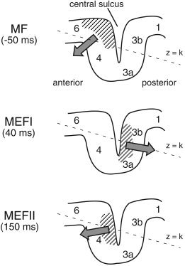
Figure 6.

Proposed generators of the motor field (MF) and first and second movement‐evoked fields (MEFI and MEFII) relative to Brodmann areas of the contralateral sensorimotor cortex. Hatched areas indicate the regions of cortex activated during each of the three peak latencies and solid arrows the approximate vector sum direction of intracellular currents. The orientation of the x–y plane of the MEG coordinate system (z = constant) is tilted in the anterior–posterior direction as shown by the dotted line. See text for further details.
