
Figure 9.
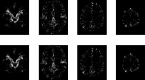
AR(1) images for face data: The top row shows estimated profiles from a tissue‐type prior (smoothed CSF versus other, prior (iii)) and the bottom row shows the estimated profiles from models with spatial GMRF priors. Columns in this figure show results for slices z = −27, 3, 33 and 63 mm.