
Figure 2.
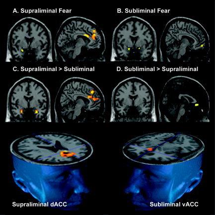
A–D: Statistical parameter maps (SPMs at P < 0.05small volume corrected), overlaid on the canonical T1 images, derived from the Montreal Neurological Institute. Images are in neurological orientation (left hemisphere = left of image). SPMs are for within‐ and between‐condition contrasts of supraliminal and subliminal fear, relative to a neutral baseline, for the regions of interest: amygdala, ventral (vACC) and dorsal anterior cingulate cortex (dACC), and connected ventral (vMPFC) and dorsal (dMPFC) portions of the medial prefrontal cortex. The within‐condition contrast of supraliminal fear relative to neutral elicited significant responses in the left amygdala (A, image on left) and left dACC/dMPFC (A, image on right). The contrast of subliminal fear relative to neutral elicited significant activity in the bilateral amygdala (B, image on left) and vACC (B, image on right). In between‐condition contrasts, supraliminal fear was distinguished by significantly greater responses in the bilateral amygdala, most pronounced in the left amygdala, and in the dACC and dMPFC (C), whereas subliminal fear was distinguished by relatively greater activity in the vACC (D). The 3D images (bottom row) illustrate further the distinction between conditions in prefrontal responses: whereas supraliminal fear was distinguished by greater responses in the dorsal ACC, supraliminal fear was distinguished by greater responses in the ventral ACC. The standardized anatomical coordinates for these regions of activity are presented in Table I.