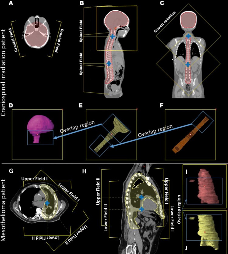
Figure 1.
Field arrangement for the craniospinal irradiation patient (A–F) and mesothelioma irradiation patient (G–J). (A) Two brain fields. (B) Upper spine field. (C) Lower spine field. (D–F) Corresponding target volumes. (G) Axial view. (H) Sagittal view. (I, J) Target volumes corresponding to the 2 upper and 2 lower fields, respectively. Field isocenters are indicated (blue cross).

