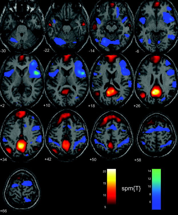
Figure 5.

Functional connectivity during rest (eyes open). Brain regions that in the eyes‐open condition correlated positively (red‐yellow color‐coded) and negatively (blue‐magenta color‐coded) with the precuneus/PCC area are matched very closely to the corresponding brain regions involved for the eyes‐closed condition (see Fig. 4 for comparison). Minor differences between the two conditions exist. For example, the positive correlation in the parahippocampal gyrus and the inferior frontal cortex was only found in the left hemisphere in the eyes‐open condition. Moreover, a negative correlation with the precuneus was found in the lateral cerebellum for the eyes‐open condition but not in the eyes‐closed condition.
