
Figure 1.
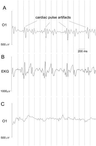
A: Raw EEG trace recorded in static magnetic field, without MRI scanning, for a representative subject (electrode O1). Peaks synchronous and delayed of a constant lag respect to cardiac activity (channel EKG) are clearly visible, indicating that they are due to cardiac pulse effect. B: Raw EKG trace recorded in static magnetic field: peaks are detected and marked. C: Reliable EEG obtained after correction procedure for electrode O1.