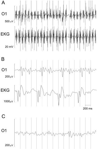
Figure 3.

A: Raw EEG traces recorded during MRI scanning for a representative subject (electrode O1 and cardiac channel EKG). During fMRI acquisition, the EEG is clearly corrupted by artifact induced by magnetic interference. B: O1 and EKG traces after application of procedure for scanning artifact removal. Peaks due to cardiac activity are visible. C: Trace at the end of the whole correction procedure.
