
Figure 4.
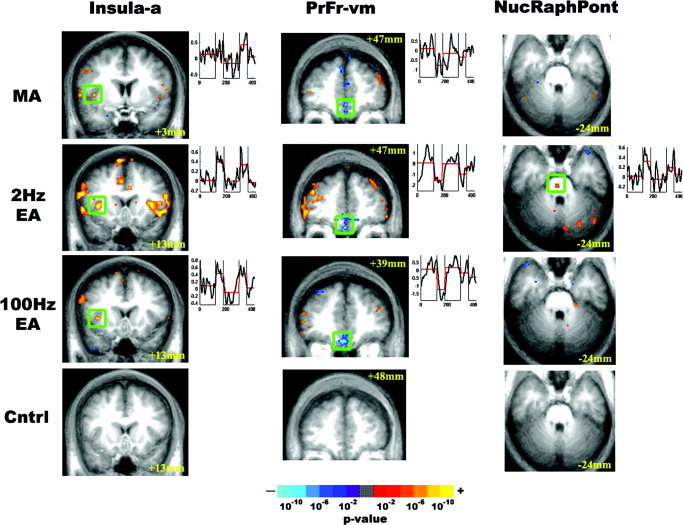
Response of limbic related structures to experimental stimulation. The results of group analysis for MA, EA, and sensory control were color‐coded based on P value and presented as activated or deactivated regions. Although all acupuncture stimulations produced signal decrease in the ventromedial prefrontal cortex and signal increase in the anterior insula, only 2‐Hz EA produced signal increase in the nucleus raphe pontis. Tactile sensory control stimulation did not produce significant response in these regions.