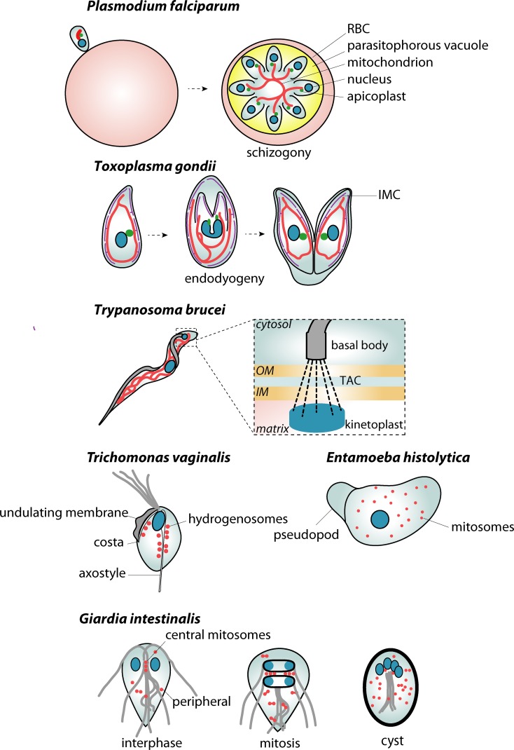
Fig 2. Key aspects of mitochondrial dynamics in parasitic protists.
Single mitochondrion of P. falciparum in synchrony with the specific cell division known as schizogony. The elongated mitochondrion branches and segregates into emerging daughter cells at the very end of the cell division. Throughout most of the cell cycle, the mitochondrion maintains the association with the apicoplast. Similarly, two daughter cells of T. gondii develop within the mother cell by a process known as endodyogeny. Here, the mitochondrion is also in contact with the IMC—a membranous compartment underneath the cytoplasmic membrane specific to Alveolata. In T. brucei, the mitochondrial genome is arranged into a kinetoplast disc, which is physically connected to the basal body of the flagellum by a TAC. The division of the single parasite mitochondrion is thus fully coordinated with the maturation of a new flagellum and the segregation of two daughter kinetoplasts. MROs of anaerobic parasitic protists are always present in higher numbers. In T. vaginalis, hydrogenosomes associate with the main cytoskeletal structures, the axostyle and costa. In G. intestinalis, central and peripheral mitosomes can be distinguished. The first are proposed to be in contact with the axonemes of flagella. Both populations divide during mitosis, which is also part of the encystation process. The mature cyst thus contains a doubled set of nuclei and mitosomes. In E. histolytica, hundreds of mitosomes are distributed all over the cell. IM, inner mitochondrial membrane; IMC, inner membrane complex; OM, outer mitochondrial membrane; MRO, mitochondrion-related organelle; RBC, red blood cell; TAC, tripartite attachment complex.

