
Figure 1.
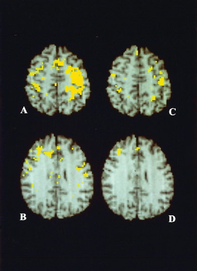
Integrated PET/MR‐images showing the anatomical location of significant mean rCBF increases in normal volunteers (hypothesis test for the mean difference in paired design; A, B), and the deficient activations in the patients with Parkinson's disease in relation to normals (hypothesis test for the difference between independent means; C, D). Activations in normal volunteers involved motor and sensory hand area, contralateral to the exploring hand, premotor cortex, supplementary motor area, superior parietal lobulus, on both sides (A) and right dorsolateral prefrontal cortex (B). Deficient activations in the Parkinson patients were at sensorimotor cortices on both sides (C), and in the Parkinson subgroup B in the right dorsolateral prefrontal and the mesial frontal cortex (D). Anatomical details are given by T1‐weighted, high resolution MR‐images of the atlas standard brain. View is from below so that the right hemisphere is seen on the left. Thresholding and correction by cluster analysis, see method section, i.e., rCBF PET—categorical comparison.