
Figure 3.
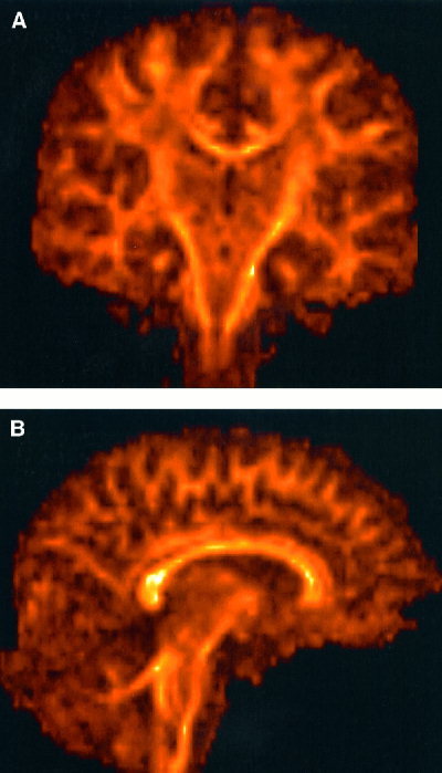
Results of reformatting the axially acquired data presented in Figure 1 into coronal plane (A) and sagittal plane (B). Note the absence of artifacts in the original slice‐selection direction (superior‐inferior), which is due to minimal slice cross‐talk resulting from a long TR.