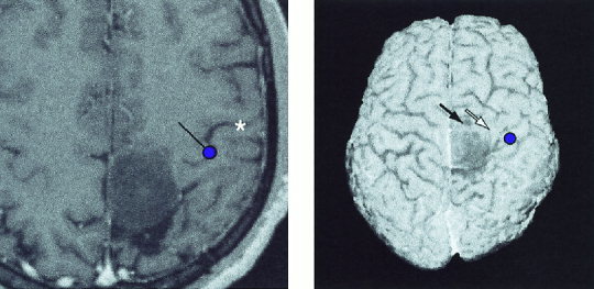
Figure 4.

Left: Source of the 20‐ms SEF to median nerve stimulation overlaid on the horizontal MR slice of Patient 5 with a recurrent parasagittal astrocytoma GII on both sides of the central sulcus. The dot gives the location and the line the current orientation. Central sulcus is marked with an asterisk. The omega‐shaped knob of the motor cortex is clearly visible. Right: N20m source to median nerve stimulation overlaid on the 3‐D surface rendering of the patient. Responses to tibial nerve stimulation were absent. In intraoperative stimulation, the motor cortex was located anterior to the tumor; stimulation of the sites pointed by black and white arrows elicited weak foot and hand movenments, respectively.
