
Figure 2.
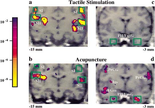
Bilateral fMRI signal increases in somatosensory cortices and signal decreases in deep structures: acupuncture needle manipulation of the left LI 4 versus tactile stimulation in a single subject. Pseudocolor KS statistical maps of signal increases (left column, a, b) and signal decreases (right column, c, d) overlaid on high‐resolution scans in gray scale at the indicated slice plane relative to the anterior commissure. Both tactile stimulation and acupuncture needle manipulation elicited signal increases in the primary and secondary somatosensory cortices (SI, SII), but the changes were more marked during tactile stimulation (a) than during acupuncture (b). Acupuncture needle manipulation elicited signal decreases in the parahippocampus/fusiform gyrus (PH/FusG) and insula (Ins) (d), whereas tactile stimulation did not (c). Also shown are acupuncture needle manipulation associated signal decreases in the middle temporal gyrus (TGm) and precentral gyrus (PrCG). Color bar shows significance, same color if signal increased or decreased.