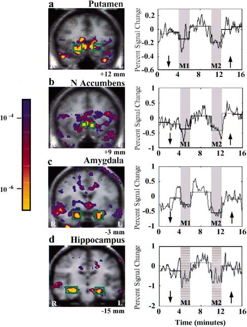
Figure 4.

Signal decreases in deep structures during acupuncture needle manipulation: group averaged data. Left LI 4 stimulation (n = 5): putamen (a), nucleus accumbens (b). Right LI 4 stimulation (n = 7): amygdala (c), hippocampus (d). The onset and recovery of signal changes showed good temporal correlation with the experimental paradigm. There was no evidence of adaptation, but rather a suggestion of enhanced effect during M2 for the amygdala (c) and the hippocampus (d).
