
Figure 3.
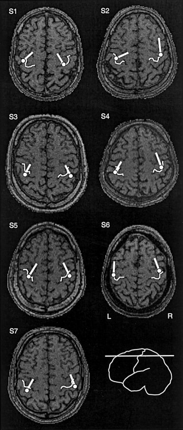
Axial MRI slices of all subjects (S1–S7) at the level of the hand area. The hand knobs are high‐lighted with white curves. The representative sources of the medial clusters and the orientations of current flow in each subject are indicated with white circles and tails. The medial clusters were typically localized in the TONGUE or ONEWORD conditions.