
Figure 1.
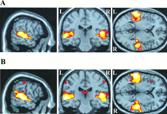
Superior temporal cortex activation during (A) passive listening to modulated tones, and (B) active listening to modulated tones, both relative to the resting baseline condition. Activation is projected onto sagittal, coronal and axial planes through the point in standard brain space corresponding to −58, −24, 4 mm in the x, y, z planes. The sagittal plane shows bilateral activation along the superior temporal gyrus and superior temporal sulcus, extending anteriorly and posteriorly beyond Heschl's gyrus. Both (A) and (B) show that the volume of temporal activation is greater on the left side in both passive and active conditions. Only active listening produced activation in other brain regions, including bilateral post‐central gyrus and left insula cortex shown here.