
Figure 5.
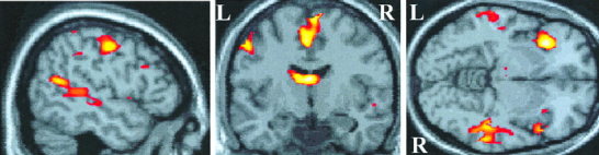
Location of preferential activation induced by the target‐detection task compared with the passive listening task. Activation is projected onto sagittal, coronal, and axial views of a canonical T1‐weighted image through the point in standard brain space corresponding to −50, −8, −6 mm in the x, y, z planes. This sagittal view shows temporal cortex activation posterior to Heschl's gyrus along the superior temporal sulcus. Activation due to active listening is also produced in other brain regions, including left pre‐central gyrus, superior frontal gyrus, thalamus, and insula cortex shown here.