
Figure 3.
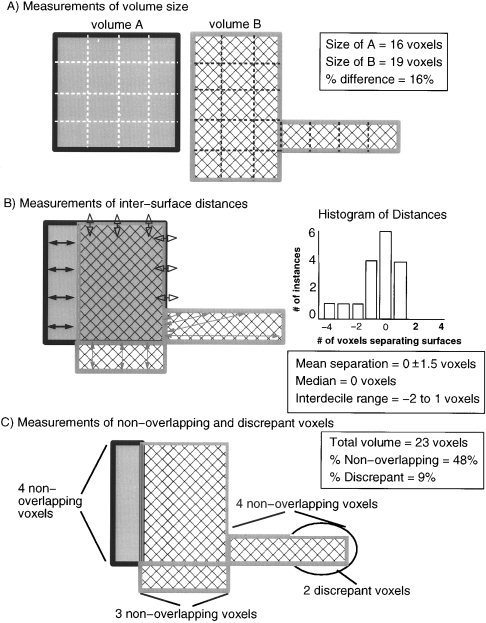
Illustration of four metrics used to compare segmented and warped lesion volumes. (A) Volume sizes were compared by computing absolute percent differences in the number of voxels included in two different segmented or warped lesion volumes. (B) Differences in the location of volumes were evaluated by measuring the relative Euclidean distances between the nearest voxels on two volume surfaces. The values computed across the surface voxels of two volumes formed a set of values that was analyzed using descriptive statistics. (C) Differences in location were also analyzed by computing the percentages of nonoverlapping and highly discrepant voxels across the total volume encompassed by a pair of segmented or warped lesion volumes.