
Figure 2.
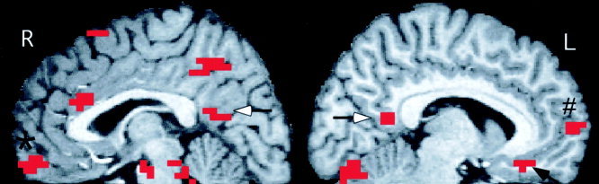
Areas of significantly greater activation in midline regions during the evaluation of pleasant compared to matched neural words. Shown are all significantly activated clusters (defined as a peak P ≤ 0. 0001 within a cluster of five voxels with P ≤ 0.005) on three adjacent sagital slices, centered at X = +3 (R) and X = −7 (L). Activations shown include the posterior cingulate cortex bilaterally (white arrows) the right anteromedial orbital cortex (*) and the left subgenual (black arrow) and the left subgenual (black arrow) and frontal polar (#) cortices. Activated voxels arer superimposed on a high‐resolution MR image normalized into Talairach space.