
Figure 2.
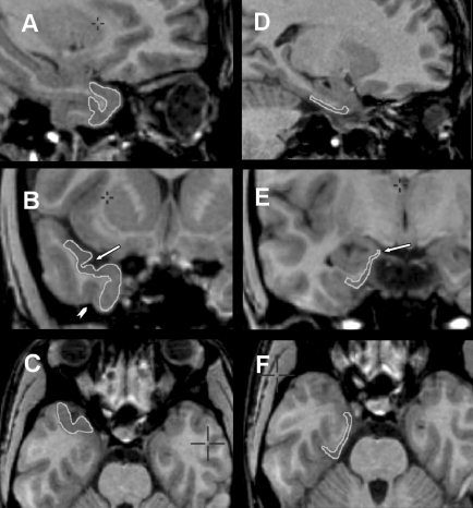
Composite of MRI showing the temporopolar cortex (A, sagittal plane; B, coronal plane; C, axial plane) and the entorhinal cortex (D, sagittal plane; E, coronal plane; F, axial plane) with anatomic boundaries delineated by edge tracing (see text for description of anatomical landmarks used for segmentation). Both the entorhinal and perirhinal cortices are constant structures, without significant changes in landmarks in the rostrocaudal axis. Arrows indicate key landmarks. B: Arrow indicates temporopolar sulcus and arrowhead indicates occipitotemporal sulcus. E: Arrow indicates the semiannularis sulcus.