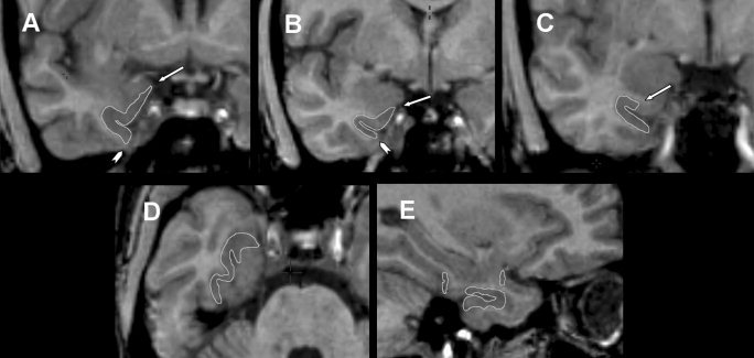
Figure 3.

Composite of MRI showing perirhinal cortex with boundaries delineated by edge tracing (A–C, coronal plane; D, axial plane; E, sagittal plane). The perirhinal cortex has significant changes in boundaries within rostrocaudal axis. A–C: Most constant aspects of the perirhinal cortex in the rostrocaudal sequence; key landmarks are labeled. A: Arrow indicates most medial point of parahippocampal gyrus and arrowhead indicates medial edge of occipitotemporal sulcus. B: Arrow indicates medial point of the medial aspect of parahippocampal gyrus and arrowhead indicates lateral edge of the lateral bank of collateral sulcus. C: Arrow indicates midpoint of the superior bank of collateral sulcus.
