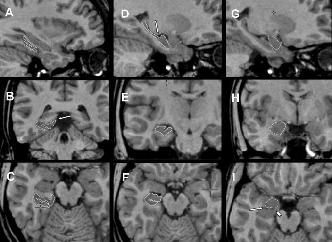
Figure 4.

Composite of MRI showing parahippocampal cortex (A, sagittal plane; B, coronal plane; C, axial plane), hippocampus (D, sagittal plane; E, coronal plane; F, axial plane) and amygdala (G, sagittal plane; H, coronal plane; I, axial plane). Parahippocampal cortex, hippocampus, and amygdala have constant boundaries (refer to text for segmentation steps) and key landmarks are labeled. B: Arrow indicates inferior limit of the inferior bank of calcarine fissure. D: Arrow indicates intralimbic gyrus closure. I: Arrow indicates temporal horn of the lateral ventricle and the arrowhead indicates the alveus.
