
Figure 3.
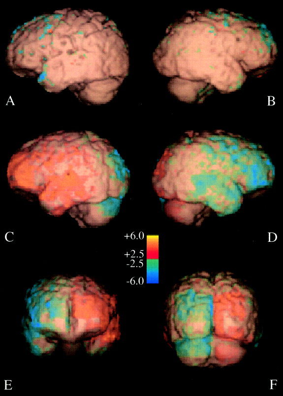
Surface rendering of a representative brain with d‐scores >2.5. The brain surface is color coded to indicate inward (blue) and outward (yellow) deformations needed to match the contralateral hemisphere. Left hemisphere colors are for Left‐to‐right analyses and right hemispheres are for Right‐to‐left analyses. (A, B) Within‐hemisphere asymmetry results with views of left and right sides. Between‐hemisphere asymmetry results with left (C), right (D), anterior (E), and posterior (F) views.