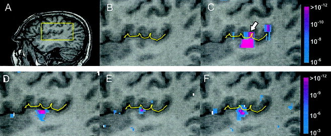
Figure 5.

Results m sixlanguage fMRI study of noa frosubject. A: Subject's sagittal anatomy with region‐of‐interest (see box) at the border of primary auditory areas and inferior parietal lobe. B: Delineation of lateral fissure. (C) Low, (D) intermediate, and (E) high spatial resolution functional maps obtained f sixdata according to the protocols described in Table II. F: Functional map obtained f sixhigh spatial resolution data that was spatially smoothed to the equivalent level of the low‐resolution data.
