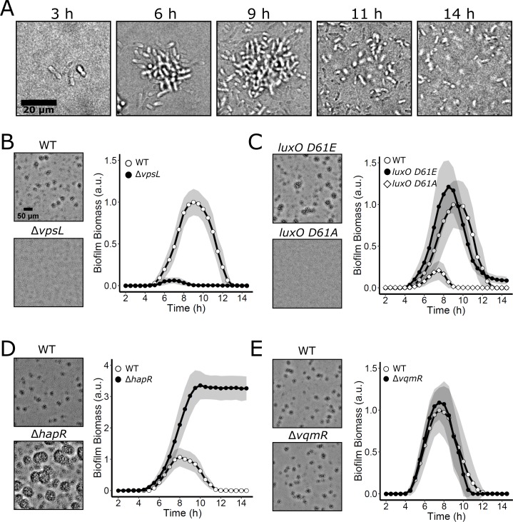
Fig 2. V. cholerae biofilm formation and dispersal under static growth conditions.
(A) Time course of a representative WT V. cholerae biofilm lifecycle as imaged by brightfield microscopy using high magnification (63× objective). (B) Left panels: brightfield projections of V. cholerae biofilms in the indicated strains after 9 h of growth at 30°C, imaged using low-magnification (10× objective). Right panel: quantitation of V. cholerae WT and ΔvpsL biofilm biomass over time. (C) As in B for V. cholerae WT and QS mutants locked in LCD (luxO D61E) and HCD (luxO D61A) modes. (D) As in B for V. cholerae WT and the LCD-locked ΔhapR strain. (E) As in B for V. cholerae WT and the ΔvqmR strain. Data are represented as means normalized to the peak biofilm biomass of the WT strain in each experiment. In all cases, n = 3 biological and n = 3 technical replicates, ± SD (shaded). Numerical data are available in S1 Data. a.u., arbitrary unit; HCD, high cell density; LCD, low cell density; QS, quorum sensing; WT, wild type.

