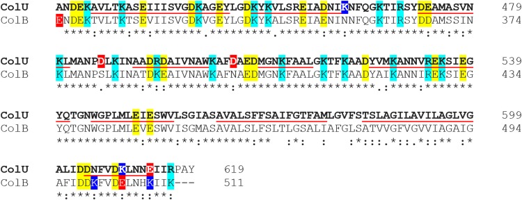
FIG 3.
Sequence alignment of C-terminal 200 amino acids of colicin U and colicin B shows high sequence similarity. Asterisks mark identical residues; colons indicate similar amino acids. Negatively charged amino acids (glutamic and aspartic acid) are in yellow; positively charged (arginine and lysine) are in cyan. Red and blue highlights denote a difference in the charge of the two sequences. The underlined sequences correspond to the expected α-helices of the pore-forming domain (69). The amino acid sequence alignment was generated with Clustal Omega (70).

