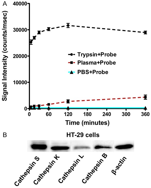
Figure 2.

Stability of cathepsin-activatable LUM015 probe in plasma and western blot analysis of cathepsin expression in colorectal cancer cells. A. Incubation of LUM015 probe with trypsin resulted in rapid activation of the probe and robust increase in fluorescence signal intensity. The signal intensity of the probe mixed with murine plasma did not significantly increase over 6 hours compare to the mixture of probe with PBS as negative control. B. Western blot analysis of the HT29 cells showed high expression of cathepsin S, B, K and to lesser degree cathepsin L.
