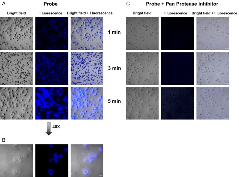
Figure 3.

In vitro assessment of LUM015 probe. A, B. Representative confocal microscopic images of incubated HT-29 cells with LUM015 shows rapid increase in fluorescence signal in the cells and extracellular space in 1-5 minutes. C. Activation of the probe was inhibited by addition of protease inhibitor cocktail to the media.
