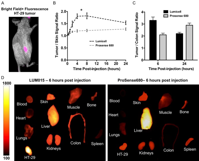
Figure 4.

Optimizing the timing for in vivo imaging of LUM015 and comparison of the biodistribution of LUM015 and Prosense680. A. Representative multispectral deconvolution fluorescence imaging of the subcutaneously implanted HT29 tumor overlaid with white light at 6 hours post LUM015 injection showed localization of the tumor with sharp margins. B. LUM015 showed overall more rapid kinetics compared to Prosense680 over 24 hours post injection with maximum tumor-to-skin ratio of 1.8±0.2 (mean ± SD) at 4-8 hours (*), and slowly decreased ratio to 1.5±0.1 at 24 hours. C. LUM015 results in maximum tumor-to-colon ratio of 3.3±0.3 at 6 hours, compared to Prosense680, which results in maximum ratio of 2.9±0.2 at 24 hours. D. Representative comparison of LUM015 and ProSense680 biodistribution at 6 hours shows more rapid clearance of LUM015 from non-targeted background tissues such as liver and spleen and higher probe accumulation in the colon tumor compared to Prosense680.
