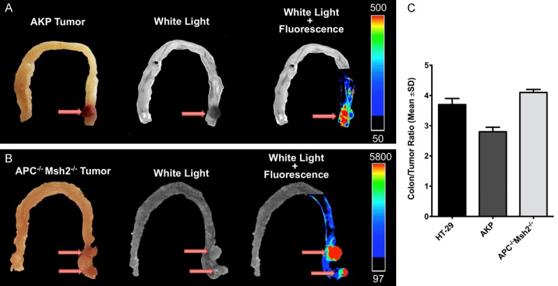
Figure 6.

Optical imaging of colorectal tumors in genetically engineered mouse models of colorectal cancer. (A) Representative optical imaging of orthotopic colon tumors in 6 hours post probe injection shows significantly increased signal intensity in the tumor relative to the normal adjacent colon tissue with tumor-to-background ratio of 2.8±0.1 in APCKOKrasLSL-G12Dp53flox/flox (AKP) tumors (A), 4.1±0.1 in APCLoxP/LoxPMsh2LoxP/LoxP tumors (B), and 3.7±0.2 in orthotopic HT-29 tumors (C).
