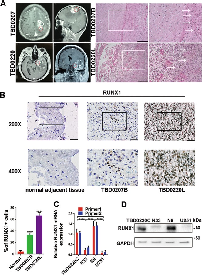
Fig. 1.
RUNX1 is upregulated in mesenchymal glioblastoma specimens and cell lines. a TBD0207B and TBD0220L represent samples that were removed from the back and left side of the tumor, respectively. Representative images of HE-stained tissues in TBD0207B and TBD0220L. Scale bar, 100 μm. b Representative tissues immunohistochemically stained for RUNX1 in normal brain tissue, TBD0207B and TBD0220L. Scale bar, 100 μm. Quantitative analysis of immunohistochemical staining for RUNX1 in a high-magnification view. n = 5 per group. c, d Western blots and qPCR were used to analyze the protein and mRNA expression levels, respectively, in TBD0220C, N33, N9, and U251 cells. GAPDH served as the negative control. (****p < 0.0001).

