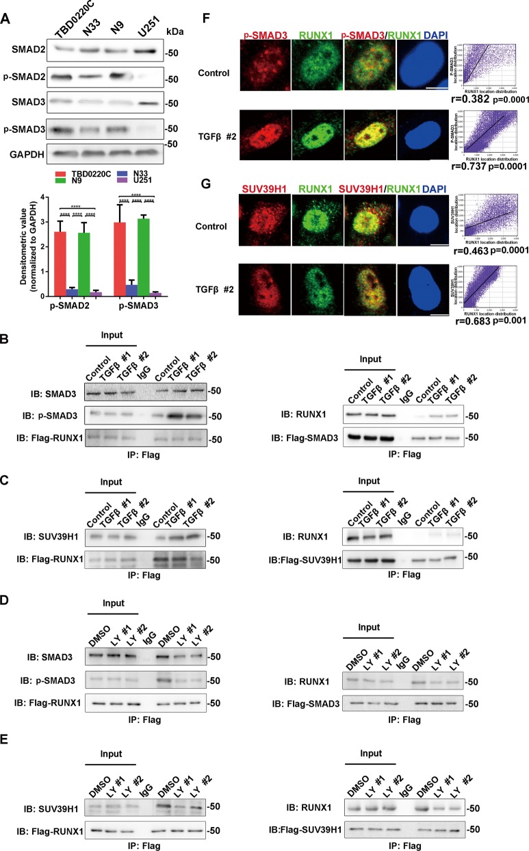
Fig. 2.
The TGFβ signaling pathway enhances the protein interaction between RUNX1 and p-SMAD3/ SUV39H1. a Western blotting analysis of SMAD2, p-SMAD2, SMAD3, and p-SMAD3 expression in four human glioma cell lines. Densitometric analysis of p-SMAD2 and p-SMAD3 is shown. n = 3 per group. b–e The interaction between RUNX1 and SUV39H1/SMAD3 was validated by co-IP. U251 cells overexpressing Flag-RUNX1 or Flag-SMAD3 (b) or Flag-SUV39H1 (c) in a background of TGFβ stimulation. N9 cells overexpressing Flag-RUNX1 or Flag-SMAD3 (d) or Flag-SUV39H1 (e) in the presence of LY2109761. f, g Subcellular localization of RUNX1, p-SMAD3 (f) and SUV39H1 (g) in U251 cells under normal or TGFβ protein conditions. Scale bar, 5 μm. A correlation analysis verified the positional overlap. n = 5 per group. (TGFβ #1 and TGFβ #2 indicate treatment of TGFβ protein for 4 h and 8 h. LY #1 and LY #2 indicate treatment of LY2109761 for 24 h and 48 h. ****p < 0.0001).

