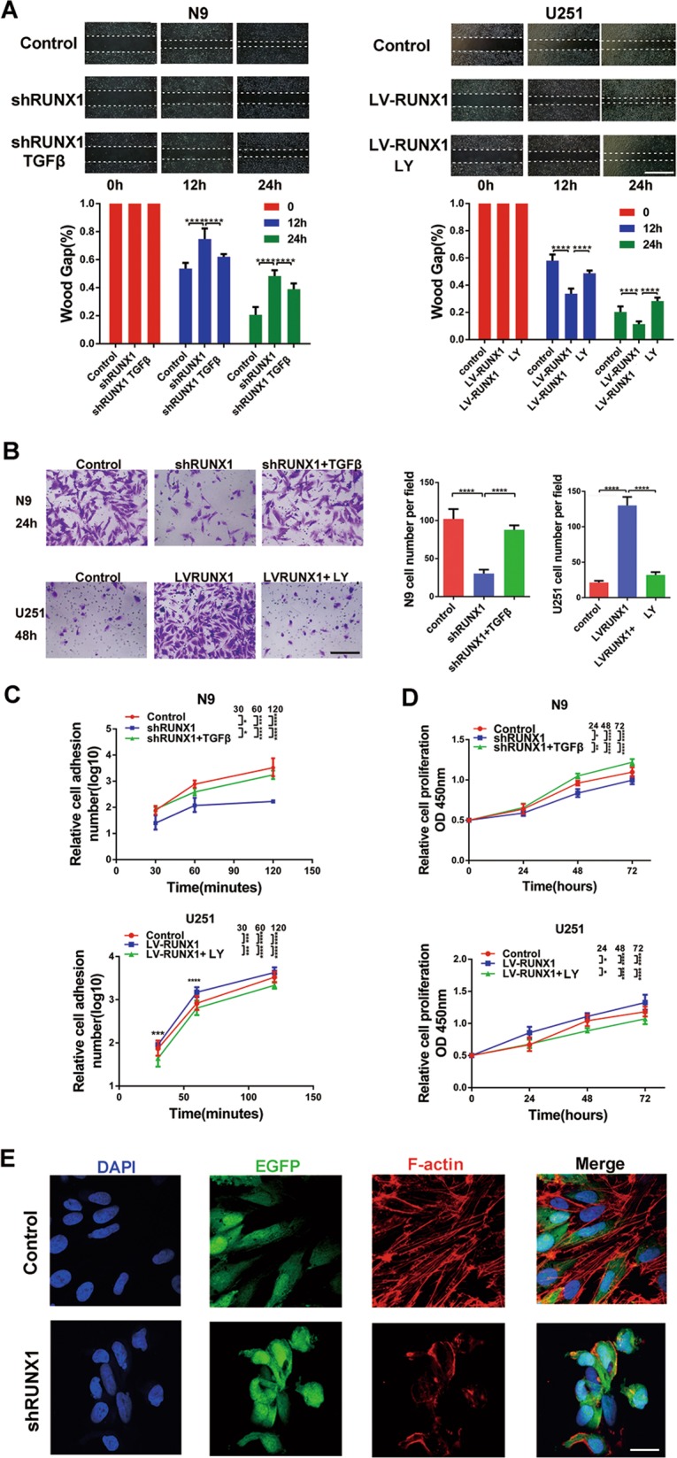
Fig. 6.

RUNX1 promotes cell proliferation, invasion and adhesion in a TGFβ pathway-dependent manner in vitro. a Wound-healing assays were used to analyze migration and invasion following treatment with shRUNX1, TGFβ plus shRUNX1, LV-RUNX1, or LY219761 plus LV-RUNX1. Scale bar, 200 μm. n = 3 per group. b N9 cells were treated with shRUNX1 or TGFβ plus shRUNX1, and U251 cells were treated with LV-RUNX1 or LY219761 plus LV-RUNX1, and Transwell assays were performed. Scale bar, 60 μm. n = 3 per group. c, d CCK8 assays and cell adhesion assays were used to analyze the proliferation and adhesion, respectively, of GBM cells. e F-actin levels were analyzed by immunofluorescence in control and shRUNX1-treated N9 cells that were transfected with EGFP lentivirus. Scale bar, 20 μm. (*p < 0.05, ***p < 0.001, and ****p < 0.0001, respectively).
