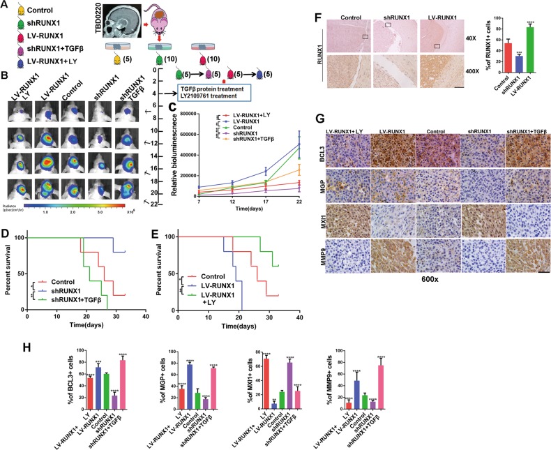
Fig. 7.
RUNX1 promotes PDX tumor proliferation and invasion in a TGFβ pathway-dependent manner in vivo. a First, nude mice were intracranially implanted with differently treated PDX tissues (control, shRUNX1, and LV-RUNX1). After 4 days LY2109761 and TGFβ were administered via the tail vein, the nude mice were divided into 5 groups, and bioluminescence imaging was performed. b Representative bioluminescence images of mice implanted with intracranial tumors on days 7, 12, 17 and 22. The relative bioluminescence values are shown (c). Survival was also measured in these 5 groups (d, e). (*, ** and *** indicate p < 0.05, p < 0.01and p < 0.001, respectively). f IHC was used to analyze the expression of RUNX1 at the control, shRUNX1 and LVRUNX1 sites.Quantitative analyses were performed using ImageJ software for each high-magnification view. n = 5 per group. g IHC staining for BCL3, MGP, MXI1 and MMP9 was performed in mouse brain slices in the control, shRUNX1, TGFβ protein plus shRUNX1, LV-RUNX1 and LY2109761 plus LV-RUNX1 groups. h Quantitative analyses of these genes were performed using ImageJ software for each high-magnification view. n = 5 per group. Scale, 20 μm. (**p < 0.01, ***p < 0.001 and ****p < 0.0001, respectively).

