
Figure 1.
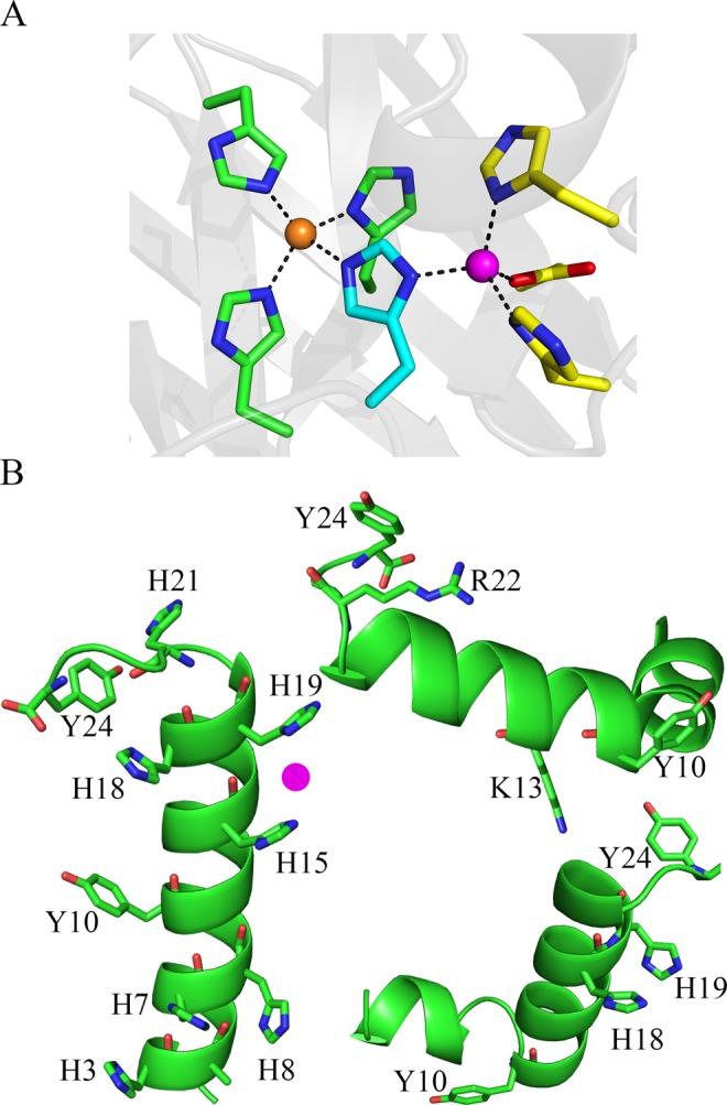
In (A), copper/zinc coordination site in superoxide dismutase (SOD) (PDB, 2SOD)57. Cu2+ (light brown sphere) is ligated by His44, His46, His61 and His118, while Zn2+ (magenta sphere) is ligated by His61, His69, His78 and Asp81. His61 (blue residue) bridges Cu2+ and Zn2+ ions. In (B), speculative PEP-FOLD models of Hst-5. Side chain labeling: left, all seven histidine and both tyrosine side chains are labeled; top, K13, R22, and both tyrosine residues; bottom right, H18, H19, and both tyrosines are labeled. The mutants, Hst-5 K13E/R22G (top) and H18A/H19A (bottom) are investigated here. A speculative binding site for the Zn2+ ion is shown in pink.