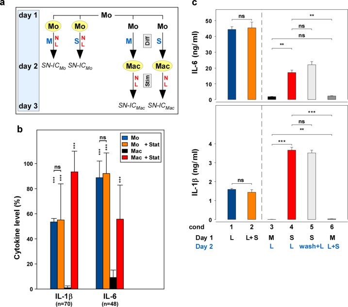
Fig. 1. Statins retain the cytokine production of monocyte-derived macrophages in the differentiation phase, but do not affect cytokine production of freshly isolated monocytes.
a Experimental design. On day 1, mononuclear cells (MNC) were prepared from heparinized whole blood. Monocytes (Mo) were isolated from MNC using CD14-antibodies linked to magnetic beads. Mo (left part of the figure; marked in yellow) were incubated in VLE-RPMI-1640, containing 10% fetal calf serum (FCS), 1% l-glutamine, and 1% antibiotics, without or with statin (10 µg/ml; M or S, respectively; blue letters) and without or with LPS (100 ng/ml; N or L, respectively; red letters), added immediately after the isolation of the Mo. On day 2, the supernatants and/or the cells were harvested and stored for analyses (SN-ICMo). On the other hand, macrophages (Mac; right part of the figure; marked in yellow) were derived from the Mo by incubating Mo without (M) or with statin (S) for 24 h (Diff, differentiation phase; gray box), but without stimulus. On day 2, medium (N) or medium with LPS (L) was added as the stimulus and the cultures were incubated for further 24 h (Stim, stimulation phase; gray box). Thereafter (on day 3), the supernatants and/or the cells of the Mac were harvested and stored for analyses (SN-ICMac). b A summary of multiple experiments shows that fluvastatin does not affect the cytokine production of the Mo, but retains the cytokine production of the Mac. Mo and Mac were isolated and prepared as described in Fig. 1a. The cytokine data of the four controls (Mo, Mo + Stat, Mac, and Mac + Stat; blue, orange, black, and red columns, respectively; all LPS-stimulated) always present in the numerous experiments performed in the study (IL-1, n = 70; IL-6, n = 48) were normalized (%) to the respective highest cytokine level of these four controls. The mean, the SD, and the significance of these data were calculated in SPSS (Levene’s test, Welch’s ANOVA, and Games–Howell post hoc analysis). The asterisks above the columns reflect the significance of “Mac” vs. “Mo”, “Mo + Stat,” or “Mac + Stat”, respectively; other comparisons are indicated by the lines (***p < 0.001; **p < 0.01; *p < 0.05; ns, not significant). c The retainment effect of the statin is initiated during the differentiation phase. Freshly isolated Mo (100,000 cells/cm2) were incubated in 24-well plates (Nunc) on day 1 and LPS (cond 1) or LPS and fluvastatin (cond 2) were added. Supernatants were harvested on day 2. In order to produce Mac cultures, the Mo were incubated on day 1 in the absence (cond 3) or presence (cond 4) of fluvastatin. On day 2, LPS was added to both cultures (blue letters). In parallel cultures to cond 4 (i.e., statin-pretreated Mac), the statin was removed by a washing step on day 2, before LPS was added (light gray columns; wash + L; cond 5). On the other hand, in parallel cultures to cond 3 (i.e., medium-pretreated Mac), LPS and statin were added simultaneously on day 2 (dark gray columns; L + S; cond 6). Supernatants were harvested after further 24 h. The cytokine concentration was determined in ELISA. Four additional experiments showed similar results. Statistics and color code as in Fig. 1b.

