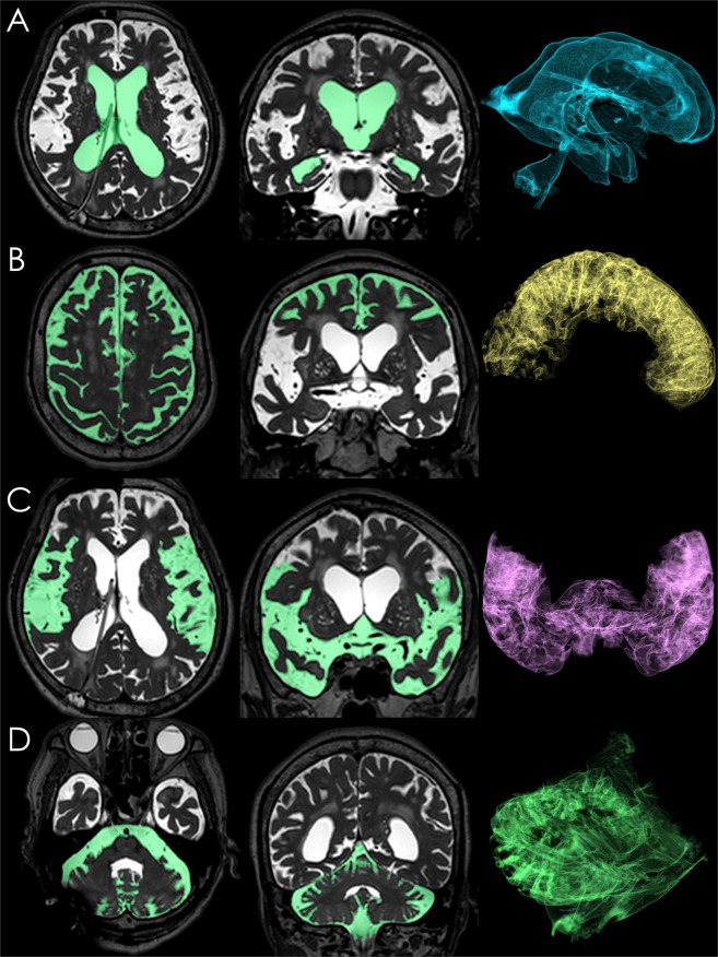
Figure 6.
Segmentation of the total ventricles and three parts of the subarachnoid spaces. These images were created on the SYNAPSE 3D workstation (Fujifilm Medical Systems, Tokyo, Japan) from a three-dimensional (3D) T2-weighted 3-Tesla sequence 10 days after ventriculoperitoneal shunting via parieto-occipital approach in a patient diagnosed with co-occurrence of idiopathic normal pressure hydrocephalus (iNPH) and Alzheimer’s disease (AD). The ventricles were manually extracted from intracranial cerebrospinal fluid (CSF) space by enclosing in a free shape. (A) The total subarachnoid spaces were segmented by subtraction of the total ventricles from total intracranial CSF spaces. The convexity subarachnoid space (B) was manually segmented from the total subarachnoid spaces per the anatomical landmarks of the basal interhemispheric cistern, Sylvian fissure, and tentorium cerebelli. From the residual subarachnoid spaces, the subarachnoid space in the posterior fossa (D) was segmented with reference to the anatomical landmarks of the tentorium cerebelli, chiasma and optic tract. There was a small MRI artefact in the right occipital posterior fossa. The Sylvian fissure and basal cistern (C) were segmented by subtraction of the convexity subarachnoid space and subarachnoid space in the posterior fossa from the total subarachnoid spaces. The convexity subarachnoid space per ventricle ratio (CVR), which was defined as the volume of the convexity subarachnoid space (178 mL) divided by the total ventricular volume (118 mL), was 1.51.

