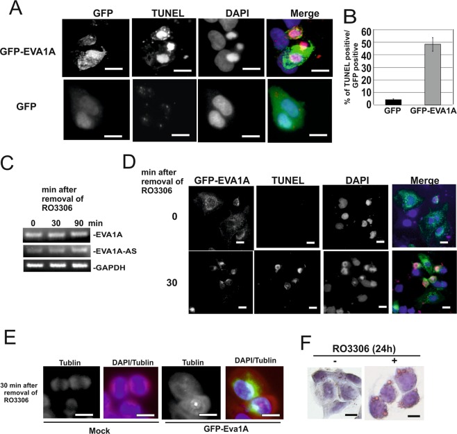
Figure 3.
Ectopic expression of EVA1A in HCC cells caused cell death. (AB) GFP-EVA1A or GFP was overexpressed in HepG2 cells and applied for TUNEL (red), and DAPI (blue) staining. Bars represent 40 µm. (A) The ratio between GFP: TUNEL positive cells was determined. (B) (C) HepG2 cells were treated with CDK1 kinase inhibitor RO3306 (5 µM) for 24 h. Total RNAs obtained from cells after removal of inhibitor (0, 30, and 120 min) were applied for EVA1A, EVA1A-AS or GAPDH specific semi quantitative RT-PCR as indicated. (D,E) HepG2 cells were transfected with GFP-EVA1A cDNA and then treated with CDK1 kinase inhibitor RO3306 (5 µM) for 24 h. Cells were fixed 0 or 30 min after removal of inhibitor. Cells were stained by DAPI and TUNEL (D) or Tubulin (E) antibody and DAPI. Three independent experiments were performed and representative images are shown. Bars represent 40 µm. (F) HepG2 cells were incubated with (+) or without (−) RO3306 for 24 h. Cells were stained by ORO. Bars represent 40 µm.

