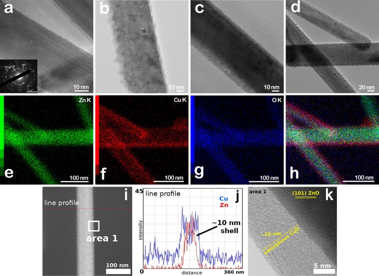
Figure 4.
TEM images of (a) ZnO nanowires having in the inset a SAED pattern proving the ZnO wurtzite phase and of (b-d) ZnO-CuxO core-shell nanowires with different shell thicknesses (with (b) ZnO-CuxO_1, (c) ZnO-CuxO_2 and (d) ZnO-CuxO_3), (e–h) Elemental maps of the same ZnO-CuxO_3 nanowires region, indicating spatially-resolved elemental distribution of Zn (green), Cu (red) O (blue), and their superposition, (i) STEM image, (j) EDS line profile analysis by STEM mode and (k) HRTEM image of area 1 of a ZnO-CuxO core-shell nanowire.

