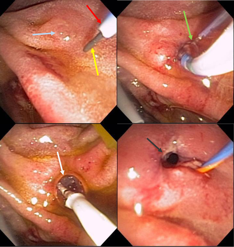
Figure 17:
Wire-guided (yellow arrow) cannulation of the minor papilla (blue arrow) with a highly tapered cannula (red arrow) followed by pull-type minor papilla sphincterotomy (green arrow) and a minor papilla endoscopic orifice dilation (white arrow). Widely open minor papilla orifice after endoscopic dilation (grey arrow).

