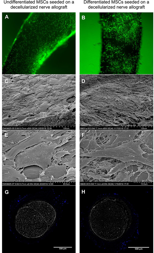
Figure 3.
Undifferentiated MSCs (left) and differentiated MSCs (right) seeded on a nerve allograft. 4a-b. Viable cells are visualized in green, dead cells in red (not present). 4c-f. Scanning Electron Microscopy images of undifferentiated MSCs seeded onto a nerve allograft (left) and differentiated MSCs seeded on a nerve allograft (right) in multiple magnifications (500X, 2000X and 3000X), showing the different morphology of the cells when seeded on the nerve allograft. 4g-h. Crosssectional images of the mid-portions of processed nerve allografts seeded with undifferentiated (left) and differentiated (right) MSCs. Cell nuclei are Hoechst-stained (bright blue). The inner ultrastructure of the nerve does not contain any cells.

