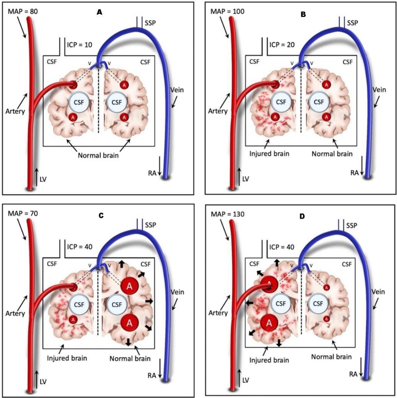
Figure 2. Hypervolemic intracranial hypertension with high and low blood pressure.
A. Normal uninjured brain with normal blood pressure and ICP
B. Injured brain (left) at a baseline ICP 20mmHg and MAP 100mmHg
C. Decreased blood pressure produces vasodilation of uninjured brain with ICP elevated to 40mmHg
D. Increased blood pressure produces vasodilation of injured brain with ICP elevated to 40 mmHg
Notably the same increase in ICP to 40mmHg associates with distinctly different physiologic conditions.
ICP, intracranial pressure; MAP, mean arterial blood pressure

