Figure 2.
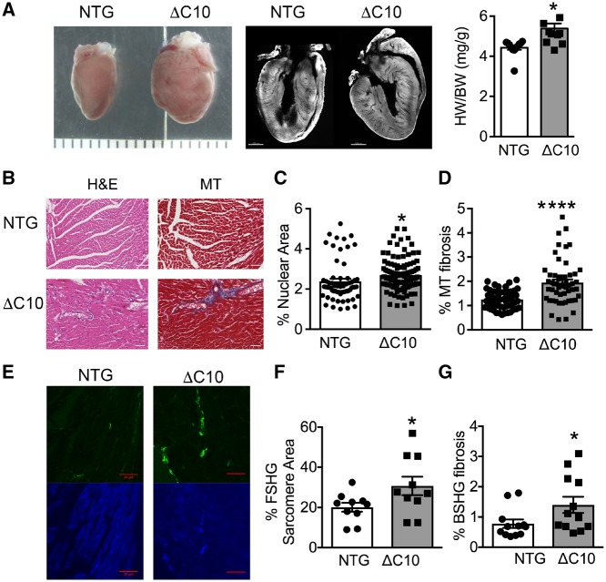
Myocardial fibrosis is elevated in cMyBP-CΔC10mut hearts. (A) Gross morphology, cross section of whole isolated hearts and heart/body weight (HW/BW) ratio (mg/g). Scale marks are separated by 1000 μm in left panel and scale bar = 1000 μm in middle panel. (B) Representative H&E- and Masson trichrome (MT)-stained myocardial sections from cMyBP-CΔC10mut and NTG hearts. Scale bar = 50 μm. (C) Quantification of percent nuclear area and (D) percent MT fibrosis obtained from measurements of H&E- and MT-labelled myocardial sections from cMyBP-CΔC10mut and NTG hearts (n = 3 NTG hearts, 62 sections and 3 cMyBP-CΔC10mut hearts, 106 sections for panel C and n = 3 NTG hearts, 53 sections and 3 cMyBP-CΔC10mut hearts, 52 sections for D). (E) Representative SHG-imaged myocardial sections from cMyBP-CΔC10mut and NTG hearts. The green channel is backward SHG (BSHG) predominantly depicting collagen fibres, and the blue channel is forward-directed SHG (FSHG) showing the sarcomere pattern. Scale bar = 20 μm. Quantification of (F) percent FSHG sarcomere area and (G) percent BSHG fibrosis obtained from SHG-imaged myocardial sections from cMyBP-CΔC10mut and NTG hearts (n = 3 NTG hearts, 10 sections and 3 cMyBP-CΔC10mut hearts, 10 sections for F and n = 3 NTG hearts, 12 sections and 3 cMyBP-CΔC10mut hearts, 13 sections for G). *P < 0.05, ****P < 0.0001.

