Figure 7.
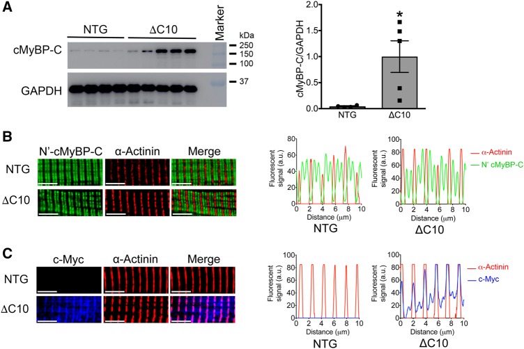
cMyBP-CΔC10mut protein is highly soluble and localizes at the Z-line in the cardiac sarcomere. (A) Western blot analysis depicting the levels of total soluble cMyBP-C (endogenous and/or cMyBP-CΔC10mut) from cMyBP-CΔC10mut and NTG hearts (n = 4 NTG, 5 cMyBP- *P < 0.027). (B) Single cardiomyocytes were isolated from cMyBP-CΔC10mut and NTG hearts and were stained with antibodies detecting cMyBP-C’s N-terminus (C0 domain) (green) and α-actinin (red). In both NTG and cMyBP-CΔC10mut myocytes, the classical staining pattern of two cMyBP-C bands between Z-lines (stained with α-actinin) was observed, indicating proper localization of endogenous cMyBP-C in cMyBP-CΔC10mut and NTG cardiomyoctyes. (C) Cardiomyocytes from cMyBP-CΔC10mut and NTG hearts were stained with antibodies to detect c-Myc (blue), which will show only the c-Myc-tagged cMyBP-CΔC10mut protein and α-actinin (red). The c-Myc signal was only observed in myocytes from cMyBP-CΔC10mut hearts. The presence of c-Myc staining at the Z-line with α-actinin suggests that the cMyBP-CΔC10mut protein aberrantly distributes within the cardiac sarcomere in cMyBP-CΔC10mut myocytes. Scale bar = 5 μm in B and C.

