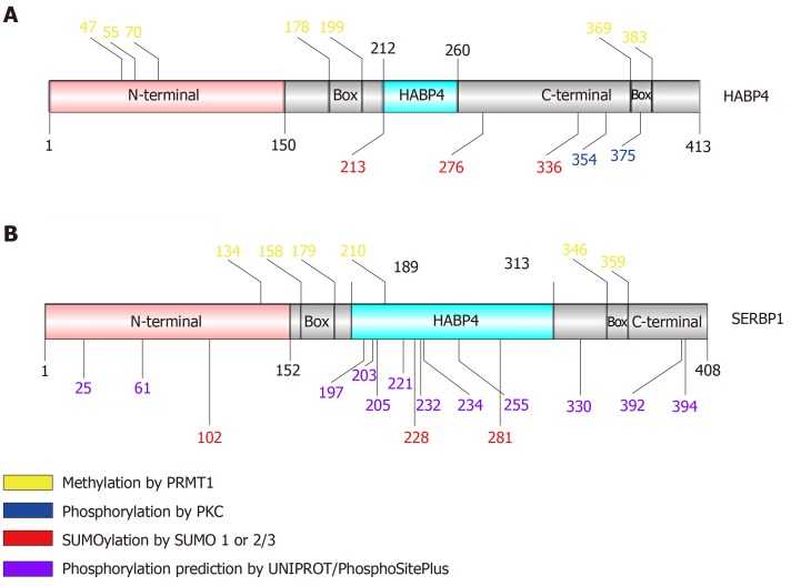
Figure 2.
Schematic view of hyaluronic acid binding protein 4 and serpin mRNA binding protein 1 primary structure identifying residues that exhibit post-translational modification. The pink region corresponds to the N-terminal domain; gray corresponds to the C-terminal; light blue corresponds to the HABP4 domain. “Box” indicates RGG/RXR boxes.

