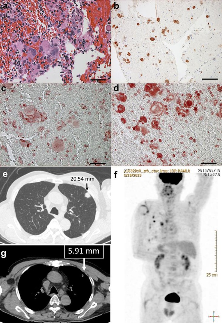
Fig. 1.
Biopsy from mediastinal lymph node: markedly atypical melanocytes (a), which were positive for S-100 (b), RANK (c) and RANKL (d). Multiple lung metastases (e), pleural, lymph node metastases, and bone metastases on PET-CT at initial visit (f). After treatment, the multiple lung metastases had decreased (g).

