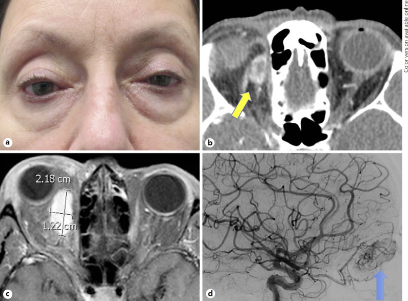
Fig. 1.
a Clinical photograph demonstrating right-sided proptosis and hypoglobus. b Orbital CT with contrast revealing an ovoid, peripherally enhancing mass at the superomedial aspect of the right orbit (yellow arrow). c Orbital MRI with contrast demonstrating a homogeneous, intensely enhancing mass within the right superomedial orbit measuring 2.2 × 1.4 × 1.3 cm. d Arterial angiography demonstrating a hypervascular mass arising from the branches of the ophthalmic artery (blue arrow).

