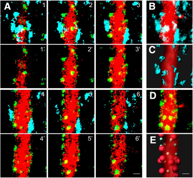
Figure 5.

FAPpost puncta on the primary apical dendrite align with presynaptic PV neurites. A, Six serial optical sections of a Pyr primary apical dendrite labeled with FAPpost (green) and dTom (red). Top row, Fluorescence aligned with presynaptic PV (YFP; cyan). Bottom row, FAPpost and dTom fluorescence alone. B, Flattened stack of the region in A, showing PV(YFP) and dTom. C, Rendering of B. D, As in B, but for FAPpost and dTom. E, Rendering of PV-assigned FAPpost puncta (large red balls) and unassigned (small green balls) puncta. Scale bar = 1 μm.
