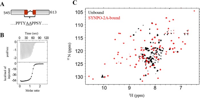
Figure 6.
SYNPO-2A binding induces changes in the KWWTD spectrum. A, schematic representation of the SYNPO-2A construct. The residues between the two PPXY motifs were replaced with two non-native alanine residues (underlined). B, ITC measurements for the KWWTD–SYNPO-2A interaction shows a binding affinity (Kd) of 0.1 μm. C, overlay of 1H-15N HSQC spectra of unbound (black) and SYNPO-2A–bound KWWTD (red) at a KWWTD:SYNPO-2A molar ratio of 1:1. Significant chemical shift dispersion in the SYNPO-2A–bound spectrum is indicative of binding-induced folding of the WW2 domain.

